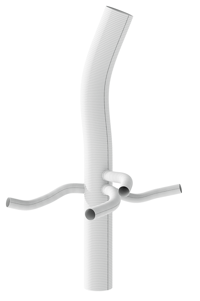
A Queensland man is now recovering well in a Chermside hospital, with full movement in his arms and legs, after undergoing one of Australia’s most complex vascular operations where surgeons used a custom 3D-printed replica of his own artery to replace a life-threateningly swollen vessel.
The Path to Recovery

Nearly three weeks after the intensive procedure, the patient has been moved out of intensive care and has begun rehabilitation. Medical staff at The Prince Charles Hospital report that he has avoided any major complications, and his strong family support has been a positive factor during his recovery. While his progress is excellent, a second follow-up surgery is planned for later this year to replace the lower portion of his aorta, addressing all residual risk from his condition.
A Ticking Time Bomb
Before the surgery, the man, who is in his late fifties, was in a perilous state. His aorta, the body’s main artery, had swelled to approximately eight centimetres—about four times its normal size.
Vascular surgeon Dr Samantha Peden explained that the artery wall was stretched so thin that a fatal rupture was imminent. The situation was described as a “ticking time bomb.” This dangerous dilation continued despite a previous surgery in 2017, leading doctors to suspect an underlying connective tissue disorder.
A Blueprint for a Breakthrough

To prepare for the high-stakes procedure, the surgical team collaborated with engineers at the nearby Herston Biofabrication Institute (HBI). The HBI team used the patient’s CT scans to create a life-sized, anatomically precise model of the damaged aorta.
The printing process took four days and used multiple materials to create a replica that mimicked the tactile feel of real tissue. This innovative model gave surgeons a hands-on understanding of the patient’s unique anatomy, allowing for meticulous planning that would not have been possible with standard 2D scans alone.
A High-Stakes Operation
The nine-hour surgery was a coordinated effort between vascular and cardiac specialists. To operate safely, they employed a technique called induced circulatory arrest, cooling the patient’s body and stopping his heart for a critical 20-minute window. Dr Peden noted that this method, while necessary, carried significant risks, including stroke or organ failure.
During this time, the surgical team removed the diseased section of the aorta and replaced it with a synthetic graft made of a flexible, waterproof fabric. This type of full aortic replacement is exceptionally complex and is performed only about six times a year at the hospital.
Published Date 22-July-2025


